QRS COMPLEXES

st segments ⇢ ⇠ p waves
ecg steps

Is the voltage normal, high, or low ?

normal

normal ventricular depolarization activity

low


weak electrical signal recorded or poor ventricular depolarization activity

(e.g. COPD, massive MI, pericardial effusion, obesity, amyloidosis)

high

strong electrical signal recorded or strong ventricular depolarization activity

(e.g. ventricular hypertrophy)

Is there ventricular hypertrophy ?


Normal ventricles: RV depolarization moves towards lead V1 (solid arrow) and away from leads V6, aVL (dashed arrows). LV depolarization moves towards leads V6, aVL (solid arrows) and away from lead V1 (dashed arrow).

Hypertrophied or enlarged RV: Large RV produces stronger depolarization hence a net electrical vector running towards V1 (strain sign and/or positive deflection of QRS complex with tall R) and away from lead V6 (negative deflection of QRS complex with deep S).

Hypertrophied or enlarged LV: Large LA produces stronger depolarization hence net electrical vector running towards V6, aVL (strain sign and/or positive deflection of QRS complex with tall R) and away from lead V1 (negative deflection of QRS complex with deep S).


Is R wave progression normal or poor?

normal

R wave starts off small and increases in amplitude going from V1 to V6. At the same time, S wave decreases in amplitude. The normal transition zone consists of isoelectric lead V3. Before the transition zone, net deflection is negative in V1-2 (small R, deep S). After the transition zone, net deflection is positive in V4-6 (tall R, small S).

poor

R wave starts off small and increases in amplitude at a slower pace and to a lesser extent going from V1 to V6. The transition zone encompasses lead V4, V5 or V6. Here, net deflection is negative in V1-4 (small R, deep S) before the transition zone, and is positive in V6 (tall R, small S) after the transition zone.

(e.g. anterior MI, left bundle branch block)

Is the QRS morphology abnormal ?

Is there bundle branch block ?

Is there fascicular block ?

Is there pathological Q wave ?

yes

wide, deep Q wave

(e.g. transmural infarct, bundle branch block, fascicular block)

dead myocardial cells (in shaded pink region) unable to produce electrical waves, thus right ventricular and septal depolarizations pull net electrical vectors away from avL and V6 resulting in the first portion (Q) of QRS complex to be negatively deflected

no

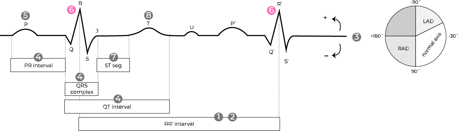
small, narrow q wave

(normal septal depolarization, often in left-sided leads, I, avL, V5-6)

myocardial cells in the ventricular septum depolarize and also pull net electrical vectors away from lateral leads avL and V6 resulting in the first portion (small q) of QRS complex to be negatively deflected

st segments ⇢ ⇠ p waves
related topics


ventricular arrhythmia
fascicular block
bundle branch block
ecg home page
myocardial infarct
pericardial disease
arrhythmia