⇧
☰
×
ECG
steps
⇲
1 | rate
2 | rhythm
3 | axis
4 | intervals
5 | p waves
6 | qrs complexes
7 | st segments
8 | t waves
specific diagnoses
⇲
acquired long qt
amiodarone toxicity
arvc/d
calcium imbalance
cardiac amyloidosis
cardiac arrest
chronic lung disease
dextrocardia
digitalis toxicity
hypertrophic cardiomyopathy
hypothermia
intracranial hemorrhage
magnesium imbalance
myocardial ischemia-infarct
localization of mi
anterior mi
inferior mi
lateral mi
posterior mi
right ventricular mi
paced rhythm
pacing modes
asynchronous pacing
single-chamber pacing
dual-chamber pacing
pericardial disease
potassium imbalance
pulmonary embolism
shock rhythm
mechanism
⇲
block
ectopy
premature
escape
tachycardia-induced ectopy
reentry
afterdepolarization
sinus
⇲
sinus node dysfunction
sinus tachycardia
atrial
⇲
premature atrial beat
atrial escape beat
atrial tachycardia
atrial flutter
atrial fibrillation
junctional
⇲
av node dysfunction
premature junctional beat
junctional escape beat
junctional tachycardia
avrt
avnrt
ventricular
⇲
bundle branch block
fascicular block
premature ventricular beat
ventricular escape beat
ventricular tachycardia
VT vs SVT
idiopathic VT
polymorphic VT
ventricular fibrillation
inherited
⇲
brugada
cpvt
early repolarization
lown-ganong-levine
long qt
short qt
wolff-parkinson-white
algorithms
⇲
arrhythmia
tachyarrhythmia
bradyarrhythmia
md doodle home
diagnosis & management
lab
cardiology
index
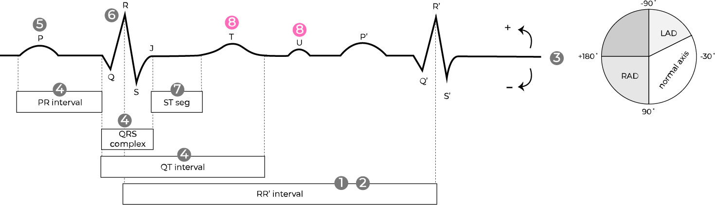
T WAVES
all steps ⇢
⇠ st segments
Are they upright or inverted ?
upright
normal, positively deflected T waves
inverted
negatively deflected T waves
(e.g. physiologic in leads V1 & aVR, lead misplacement,
myocardial ischemia
,
ventricular hypertrophy
,
hypokalemia
,
intracranial hemorrhage
,
bundle branch block
,
fascicular block
, cardiac memory, stress-induced cardiomyopathy)
Are they tall or peaked ?
yes
high T wave amplitude
(e.g.
acute phase of STEMI
,
hyperkalemia
,
benign early repolarization
)
no
normal T wave amplitude
Are there abnormal U waves ?
yes
U waves are tall or prominent
> 1/4 the height of the preceding T wave
(e.g.
sinus bradycardia
,
hypokalemia
)
no
normal U waves are upright and small
< 1/4 the height of the preceding T wave
U waves are inverted
(e.g.
myocardial ischemia
)
all steps ⇢
⇠ st segments
related topics
myocardial ischemia
hypertrophy
bundle branch block
ecg home page
potassium imbalance
ich
specific diagnoses